
Dr.RIDHAM NANDA
Abstract
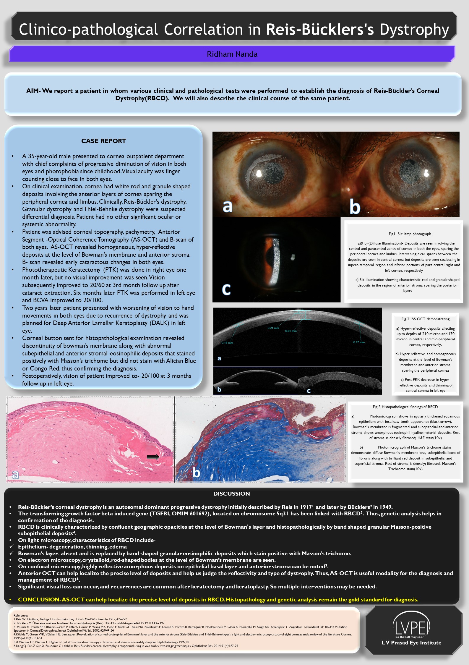
Aim- We report a patient in whom various clinical and pathological tests were performed to establish the diagnosis of Reis Bucklers Dystrophy.
Case report-A 35-year-old male presented with visual acuity reduced to 20/400 in both eyes. On clinical examination, white rod and granule shaped deposits involving the anterior layers of the cornea were noted. Anterior segment-OCT revealed homogeneous, hyper-reflective deposits at the level of Bowman’s membrane and anterior stroma. Phototherapeutic keratectomy (PTK) was done in both the eyes. Two years later, patient presented with recurrence and was planned for deep anterior lamellar keratoplasty. Corneal button sent for histopathological examination revealed abnormal subepithelial and anterior stromal eosinophilic deposits deposits that stained positively with Masson’s trichome, thus confirming the diagnosis.
Conclusion- AS-OCT can help localize the precise level of deposits. Histopathology remains the gold standard for diagnosis of RBCD.
Leave a Comment