
Dr.AARUSHI SAINI
Dr. JOLLY ROHATGI, Dr.ANCHAL ARORA, Dr.ISHA ACHARYA
Abstract
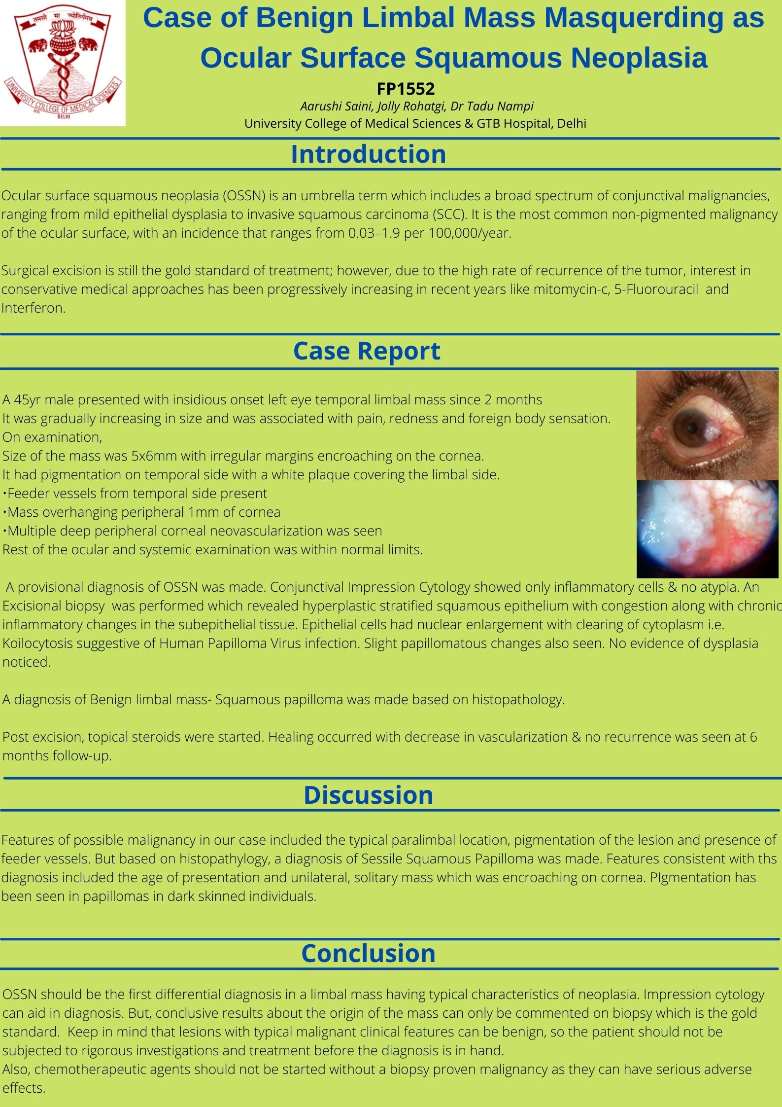
A 45yr M presented with progressively increasing, fleshy,elevated & vascularised mass on temporal limbal in LE since 2months, with complaints of foreign body sensation & watering. Mass was 5x6mm with irregular margins encroaching on the cornea. It was pigmented on the temporal side, & a white plaque covered the limbal side. Multiple & deep vascularisation of adjoining peripheral cornea was seen, a dellen was also present. Rest examination was WNL. Provisional diagnosis of OSSN was made. Impression Cytology showed only inflammatory cells & no atypia. Excisional biopsy revealed hyperplastic stratified squamous epithelium with unremarkable subepithelial tissue with no evidence of dysplasia. Post excision, topical steroids were started. Healing occurred with decrease in vascularization & no recurrence was seen at 3months follow-up.
Suspicious looking limbal mass should have OSSN as a first differential diagnosis at this age. However, biopsy is the gold standard for confirming diagnosis.
Leave a Comment