
DR. ZULAIKHA V
Dr. PADMAMALINI MAHENDRA DAS, Dr.Ankush Kawali, Dr. SANJAY SRINIVASAN, Dr.Shetty Bhujang K
Abstract
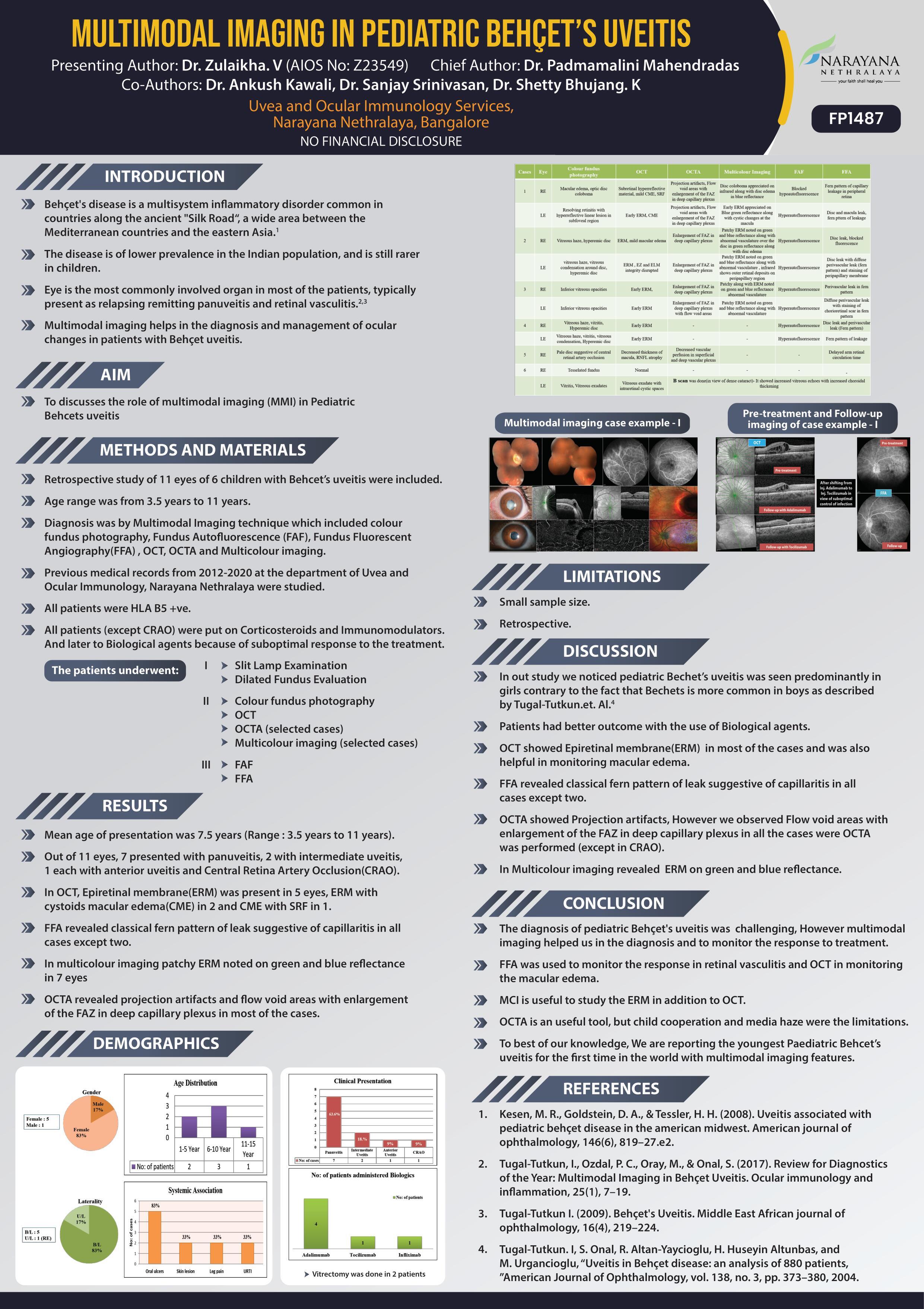
AIM: To discusses the role of multimodal imaging(MMI) in Pediatric Behcets uveitis
METHODS: Retrospective observational study of 11 eyes of 6 children with Behcet uveitis were studied using MMI including colour fundus photography, Fundus Fluorescent Angiography(FFA), OCT in all cases and OCTA in selected cases.
RESULTS: Mean age of presentation was 7.5 years.Out of 11 eyes, 7 presented with panuveitis,2 with intermediate uveitis,1 each with anterior uveitis and Central Retina Artery Occlusion(CRAO). In OCT, Epiretinal membrane(ERM) was present in 3 eyes, ERM with cystoids macular edema(CME) in 2 and CME with SRF in 1. FFA reveaed classical fern pattern of leak suggestive of capillaritis in all cases except one.In multicolour imaging patchy ERM noted on green and blue reflectancein 3 eyes.OCTA revealed enargement of FAZ in deep capillary plexus in 3 eyes.
CONCLUSION: MMI can be used in the diagnosis and to monitor the response to treatment in Paediatric Behcet’s uveitis
Leave a Comment