
Dr.ANKITA GOEL
Dr. EVA RANI TIRKEY, Dr. SHASHI AGARWAL, Dr. SUJATA LAKHTAKIA
Abstract
AIM: To report a typical case of F.K. syndrome due to rare bifrontal parafalcine meningioma.
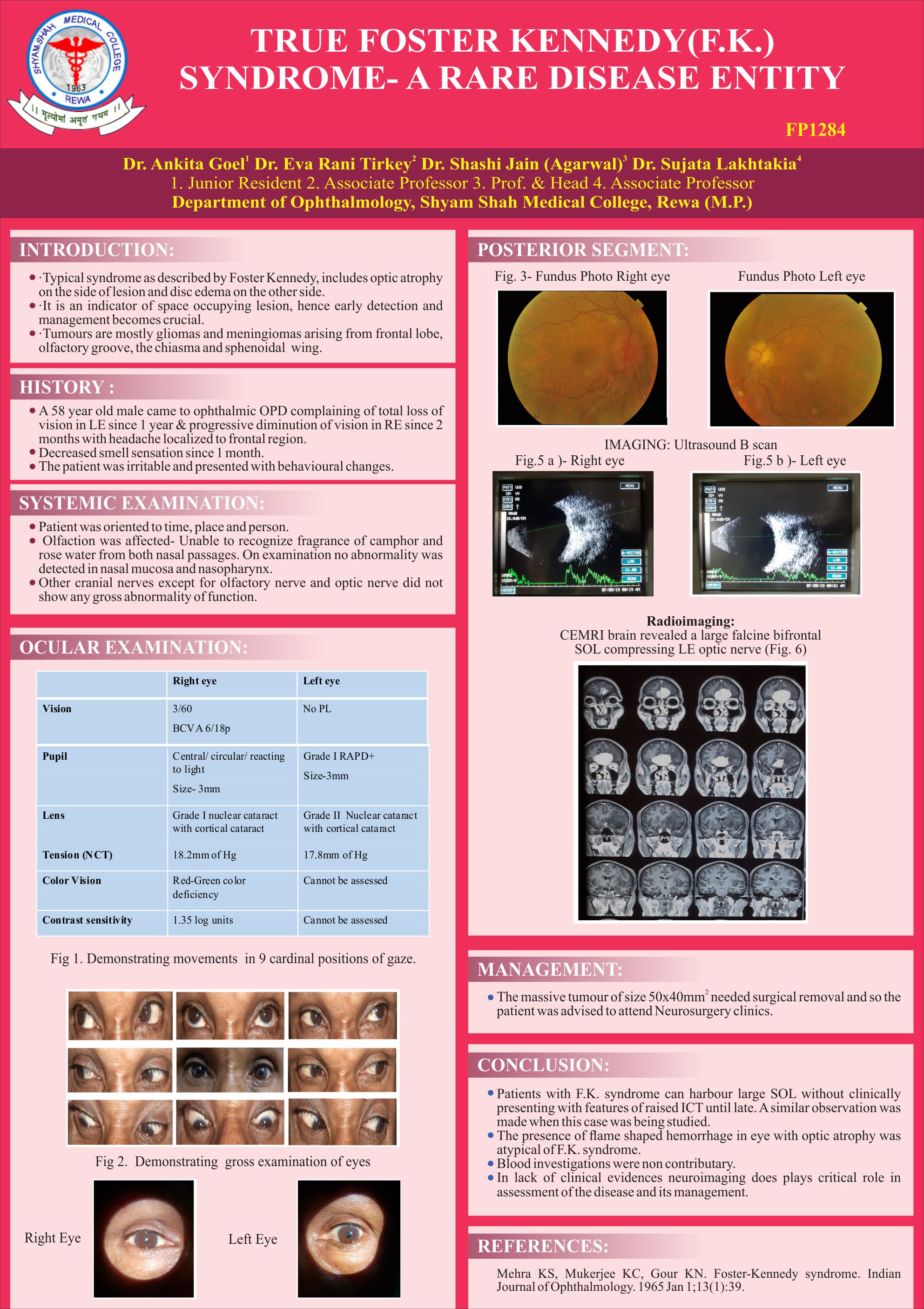
METHOD: A 58 year old male came to OPD complaining of total loss of vision in LE & progressive DOV in RE since 1 year & 2 months respectively with headache localized to frontal region & irritable behaviour. Cranial nerves examination noted defective olfaction from B/L nostril in absence of nasal abnormality. Ophthalmic examination noted VA- RE 3/60, LE No PL; LE RAPD; Red green color defect in RE; Fundus RE- Frisens Grade III papilloedema & LE pale disc with flame shaped hemorrhage. CEMRI brain revealed a large falcine bifrontal SOL compressing LE optic nerve.
RESULT: Prior to CEMRI NAION RE with LE post neuritic or compressive optic neuropathy; IIH were differentials other than F.K. syndrome.
CONCLUSION: Patient with F.K. syndrome with large SOL can be stable without any symptoms of raised ICT. In lack of clinical evidences neuroimaging plays critical role essential for patient management.
Leave a Comment