
DR. AVANTIKA SRIVASTAVA
Dr. RADHAKRISHNA MANDAL, DR. AVIK DEY SARKAR, Dr. SANJAY KUMAR DAULAT THAKUR
Abstract
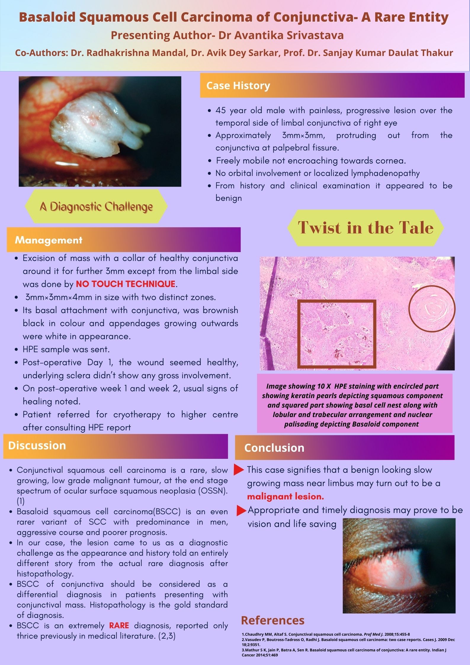
Case Presentation: A 45 year old Male presented with a 3 mm x 3 mm painless, slow growing, gelatinous, freely mobile, benign looking mass on temporal side of limbus in the conjunctiva of right eye for 1 year. Other ophthalmic findings were unremarkable. Orbital CT showed no orbital involvement or localized lymphadenopathy. The mass was excised with a healthy collar of 3 mm and was sent for Histopathological Examination (HPE). The patient was followed up with usual signs of healing. The case took a surprising turn when HPE showed areas of Keratin Pearls of squamous origin along with lobules of malignant cells displaying Nuclear palisading and Basal cell nests thereby diagnosing Basaloid Squamous Cell Carcinoma of Conjunctiva. Positive staining for 34βE12 and EMA on Immunohistochemistry confirmed it. Conclusion: Identification is highly important because of its aggressive course with frequent recurrences and distant metastasis. It has been reported only thrice before in literature.
Leave a Comment