
Dr.AAYESHA KHANUM
Dr. THIRUMALESH M. B., Dr. JIVITESH SINGH
Abstract
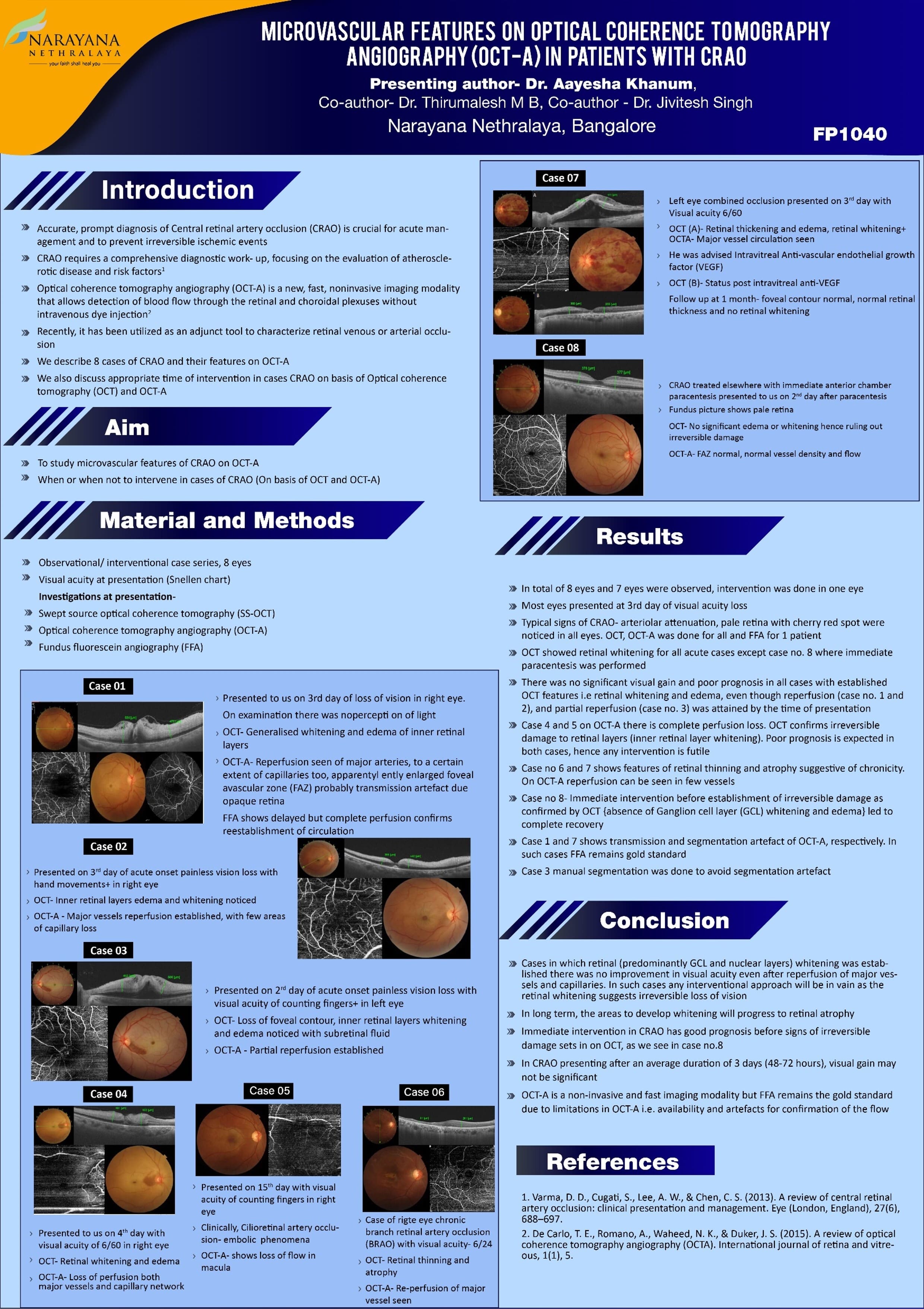
Aim: To elucidate microvascular perfusion in patients with Central retinal artery occlusion (CRAO) using OCT-A.
Material and Methods: Patients who presented to us with Central retinal artery occlusion were included in the study. OCT and OCT-A were done. Perfusion was studied using OCT-A and anatomical changes were noted in each layer. Where feasible, it was compared with Fundus fluorescein angiography (FFA).
Results: Significant intraretinal anatomical changes in different layer were noted in form of disorganization of intraretinal vasculature, hyper reflectivity of ganglion cell layer, disruption of myoid and ellipsoid zone. Significant microvascular hypoperfusion noted in both superficial and deep capillary layer, which were better demonstrated in OCT-A than FFA. We were able to differentiate between CRAO caused by either occlusive or spasmodic event.
Conclusion: OCT-A is a better and non invasive modality to demonstrate perfusion and anatomical changes in macula than FFA.
Leave a Comment