
Dr. PRAGN MEHTA
Dr. RAJESH RAMANJULU, Dr. MAHESH P. SHANMUGAM, DR.DEVASHISH DUBEY, Dr. DIVYANSH MISHRA
Abstract
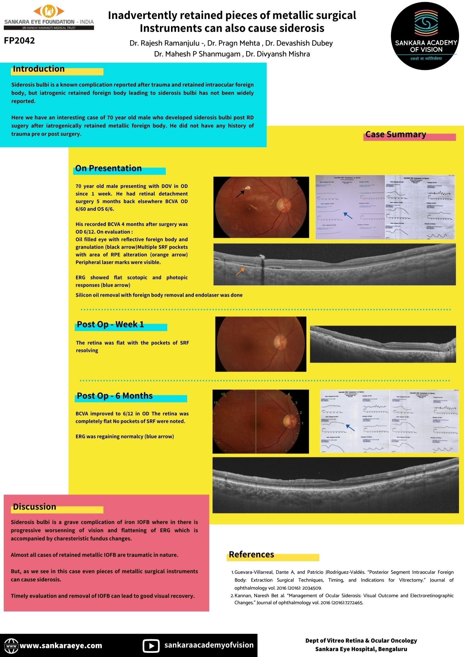
A 70-year-old male presented with blurring of vision in the right eye for 7 days. He had a history of undergoing retinal detachment surgery 5 months back elsewhere. He had no history of ocular trauma prior to or after the surgery. BCVA was OD 6/60 and OS 6/6. Intra ocular pressure was within normal limits OU.
On fundus evaluation globe was oil filled with a white reflective foreign body (FB) with surrounding granulation tissue near the superior arcade. Multiple SRF pockets with area of RPE alteration were present around to the FB. Peripheral laser marks were visible. ERG showed flat scotopic and photopic responses.
SOR with IOFB removal with EL was done.
5 months post surgery, the retina was flat with ERG responses regaining normalcy. His recorded BCVA was OD 6/12.
Conclusion : Siderosis bulbi is a grave complication of iron IOFB and even pieces of metallic surgical instruments can cause siderosis. Timely evaluation and management can lead to good visual recovery.
Leave a Comment